Case study of JF
Female, Caucasian, born 1961
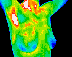
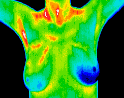
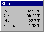
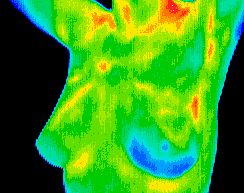
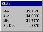
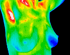
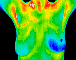
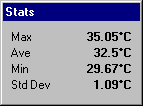
JF presented for a full body study 30-August, amongst other physical complaints which were the motivating factor for the imaging session, JF had a Hx of right sided breast cancer which was in the six o’clock lower section of the breast tissue. She had a negative result returned on an ultrasonic examination in Feb.,1998; a lumpectomy June, 1998 which returned a positive pathology result (malignant grade 3). Chemotherapy and radiation treatment followed. Her breast images on initial presentation show a temperature differential of just under 2.0OC. Vascular patterns consistent with the hyperthermic asymmetry were also noted.
JF had a scheduled appointment with her surgeon for a check up a few days after this scan, so no specific recommendation was made other than the request for her specialist to examine the right breast (which would have occurred anyway). He was satisfied that the breast tissue was without risk, so JF was recommended to return for a follow up scan in three months.
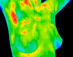
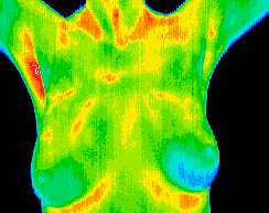
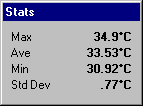
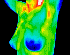
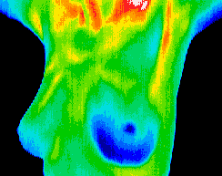
Above – This series was taken on the 11th of November, 1999, and reveals a reducing visible vascular pattern and reducing temperature asymmetry, with a difference of <1.0OC average temperature Below – The last series was taken on 6th of March, 2000, and shows the reduction in the visible vascularity in line with the previously noted reductions.
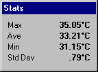
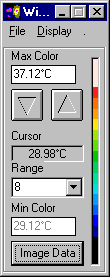
With the average temperature values varying <0.8OC, and the vascular patterns still visibly receding, the efficacy of JF’s treatment regime is confirmed, and future monitoring of her breast’s vascular supply will be undertaken.
Conclusion
This study demonstrates the ability of correct Thermographic techniques to assess the patient’s response to a treatment regime. In cases like this, where the specialist and physician of record do not wish to expose a patient to any further ionising radiation, Thermography offers a viable imaging modality that supplies objective, reliable results. Angiogenesis is a primary marker of breast disease which is increasing in significance following recent and ongoing research. Thermography is ideally placed as a technology to provide very early detection of an angiogenic process, as well as tracking the reduction on the vascular activity in a post therapy situation.